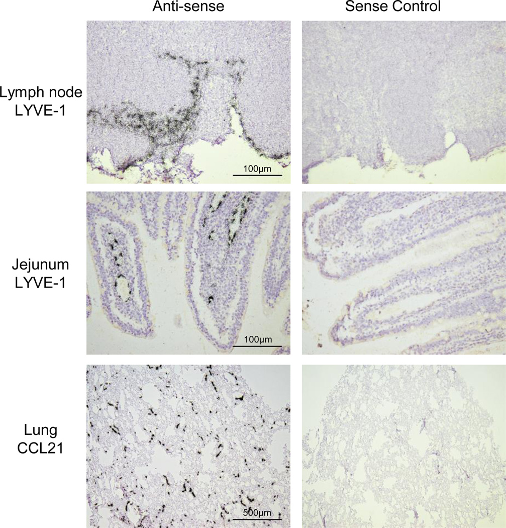
Fig. 6.
In situ hybridization localization of lymphatic marker mRNAs in ferret ferret lymph node, jejunal and lung tissues. 35S-UTP-labeled riboprobes specific for ferret LYVE-1 or CCL21 were generated and used localize cells expressing the respective mRNAs in the indicated tissue from normal ferrets. ISH signal is visualized by collections black silver grains over cells. Parallel ISHs were performed with the cognate sense control probe (micrographs on the right). Exposure times were 21d.

