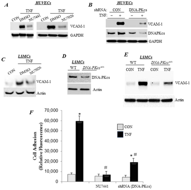
Figure 1. DNA-PK protein level and function are required for VCAM-1 expression and adhesion of inflammatory cells to endothelial cells upon TNF-α treatment.
(A) HUVECs were pretreated with 1 μM NU7026 or NU7441 or 0.1% DMSO for 30 min before treatment with TNF-α (TNF) for 24 h in the continued presence of the drugs. Protein extracts were subjected to immunoblot analysis with antibodies to VCAM-1 or GAPDH. (B) HUVECs were transduced with a lentiviral vector encoding control or shRNA targeting DNA-PKcs. Cells were then treated with TNF-α for 24 h, after which protein extracts were subjected to immunoblot analysis with antibodies to VCAM-1, DNA-PKcs, or GAPDH. (C) LSMCs were pretreated with NU7026 or 0.1% DMSO for 30 min before treatment with TNF-α for 24 h in the continued presence of the drugs. Protein extracts were subjected to immunoblot analysis with antibodies to VCAM-1 or actin. (D) Immunoblot analysis of protein extracts from WT or DNA-PKcs+/− LSMCs with antibodies to DNA-PKcs or actin. (E) WT or DNA-PKcs+/− LSMCs were treated with TNF-α for 24 h. VCAM-1 protein expression was assessed by immunoblot analysis. (F) HUVECs, subjected to DNA-PKcs knockdown or treated with NU7441, were stimulated with TNF-α for 18 h before the addition of calcein-AM-labeled U937 cells. Non-adherent monocytes were removed, and fluorescence was measured. Fluorescence is expressed in arbitrary units. *, difference from untreated cells; #, difference from TNF-α-treated cells; p<0.01.

