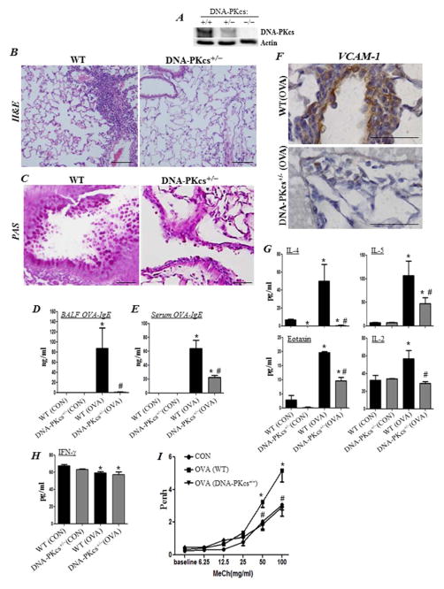
Figure 4. DNA-PKcs heterozygosity is sufficient to reduce asthma-associated traits.
WT and DNA-PKcs+/− mice were subjected to OVA sensitization followed by challenge or left untreated as described above. (A) Immunoblot analysis of lung tissue with antibodies to DNA-PKcs or actin. H&E (B) and PAS (C) staining of lung sections. Assessment of BALF (D) or sera (E) collected from the different experimental groups for OVA-specific IgE; *, difference from unchallenged mice; #, difference from OVA-challenged WT mice; p < 0.01. (F) IHC staining of lung sections with antibodies to mouse VCAM-1; bars: 5 μm. BALF from the different experimental groups were assessed for IL-4, IL-5, eotaxin, IL-2 (G) or IFN-γ (H). (I) Assessment of Penh using whole body plethysmography. Results are plotted as maximal fold increase of Penh relative to baseline and expressed as mean ± SEM where n=6 mice per group. *, difference from control WT mice; #, difference from OVA-challenged WT mice, p < 0.01.

