
Figure 3. Different proteolytic stability of the ACHM-linked mutants of PDE6C.
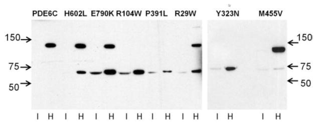
EGFP-PDE6C and mutant PDE6C proteins were extracted from transgenic eyeballs with isotonic (I) and hypotonic buffer (H) and analyzed by immunoblotting with anti-GFP antibody. The full-length EGFP-PDE6C and mutants correspond to the ~130 kDa band. The ~70 kDa band represents an N-terminal proteolytic fragment of PDE6C fused to EGFP.