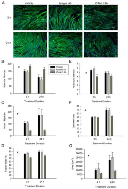
Figure 6.
The extracellular domain of ICAM-1 in the regulation of myotube fusion, alignment and size. ICAM-1+ cells were treated with vehicle, isotype control antibody (Isotype Ab;100 μg/ml), or ICAM-1 antibody (ICAM-1 Ab;100 μg/ml) at 5 d of differentiation for 2 or 24 h. A) Representative images of myosin heavy chain (green) and nuclei (blue) in ICAM-1+ cells after 2 and 24 h treatment with vehicle or antibody (scale bar = 100 μm). Quantitative analysis of myotube number (B), average number of nuclei within myotubes (C), and fusion index (D), as well as myotube alignment (E), diameter (F), and area (G) (n=4). # = different for ICAM-1 Ab compared to Isotype-Ab and vehicle (main effect for treatment; p < 0.05).

