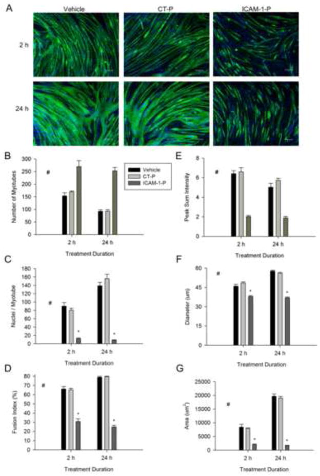
Figure 7.
The cytoplasmic domain of ICAM-1 in the regulation of myotube fusion, alignment and size. ICAM-1+ cells were treated with vehicle, control peptide (CT-P; 50 μg/ml) or ICAM-1 peptide (ICAM-1-P; 50 μg/ml) at 5 d of differentiation and myotube indices were quantified 2 and 24 h after treatment. A) Representative images of myosin heavy chain (green) and nuclei (blue) in ICAM-1+ cells after 2 and 24 h of treatment with vehicle, CT-P, and ICAM-1-P (scale bar = 100 μm). Quantitative analysis of myotube number (B), average number of nuclei within myotubes (C), and fusion index (D), as well as myotube alignment (E), diameter (F), and area (G) (n=4). # = different for ICAM-1-P compared to CT-P and vehicle (main effect for treatment; p < 0.001), * = lower for ICAM-1-P compared to CT-P and vehicle at indicated duration of treatment (interaction effect; p < 0.001).

