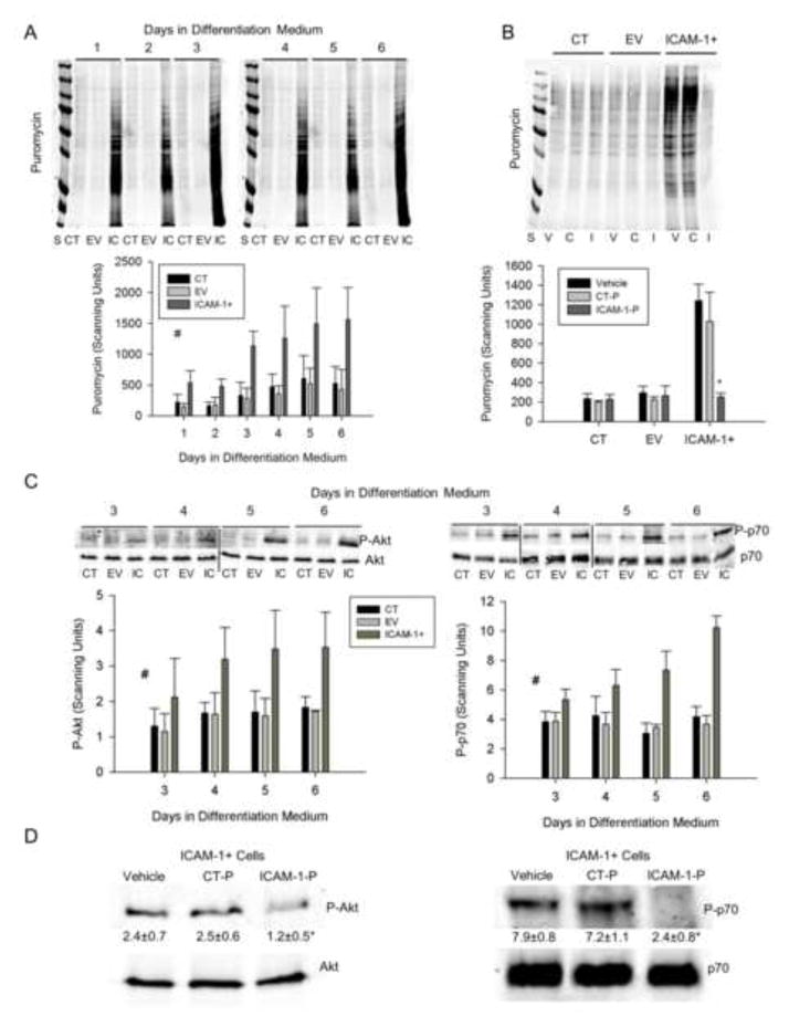
Figure 8.
Protein synthesis and Akt/p70s6k signaling. A) Representative western blot and quantitative analysis (n=4) of puromycin incorporation into nascent proteins of cells treated with differentiation medium for up to 6 d (25 μg/lane). S = standards (250-15 kDa), CT = control, EV = empty vector, IC = ICAM-1+. # = higher for ICAM-1+ compared to CT and EV cells throughout 6 d of differentiation (main effect for cell line; p < 0.001). B) Representative western blot and quantitative analysis (n=4) of puromycin in CT, EV and ICAM-1+ cells treated with vehicle (V), control peptide (C or CT-P; 50 μg/ml), or ICAM-1 peptide (I or ICAM-1-P; 50 μg/ml) at 5 d of differentiation for 2 h prior to collection of cell lysates. * = ICAM-1 peptide reduced protein synthesis in ICAM-1+ cells to levels that were observed in CT and EV cells (interaction effect; p < 0.001). C) Representative western blots of Akt and p70s6k, as well as quantitative analysis of phosphorylated Akt (Ser473; P-Akt) and p70s6k (Thr389; P-p70) at 3–6 d of differentiation. # = higher for ICAM-1+ compared to CT and EV cells (main effect for cell line; p < 0.005). D) Representative western blots of Akt and p70s6k, as well as quantitative analysis of phosphorylated levels Akt and p70s6k for ICAM-1+ cells treated with vehicle, control peptide (CT-P; 50 μg/ml) or ICAM-1 peptide (ICAM-1-P; 50 μg/ml) at 5 d of differentiation for 2 h prior to collection of cell lysates. Scanning units are presented below blots for P-Akt and P-p70. * = lower for ICAM-1-P compared to vehicle and CT-P (p < 0.05).

