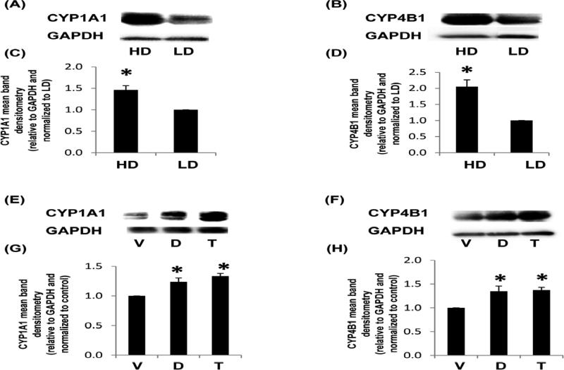
Fig. 4. CYP1A1 and CYP4B1 production from MCF10A cells in response to density or ligands known to activate HRE and XRE.
Cells were cultured in low density (LD) or high density collagen gels for 24 h and assessed for P450 enzyme production (A-D) or cultured in LD collagen gels +/− vehicle control (V), 100 μM DFOM (D) or tranilast (T) for 24 h (E-H). Representative immunoblots are displayed (A,B,E,F) and the mean band densitometry relative to the GAPDH and normalized to the control are charted (C,D,G,H) +/− SEM, N>4 *p<0.05 vs respective control.

