Figure 1.

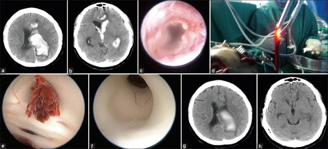
(a) Massive left lateral and paraventricular hemorrhage secondary to arteriovenous malformation. (b) The hemorrhage extends to the third ventricle. (c) The inner cortex is sometimes used to guide the surgeon. (d) Blood clot is removed. (e) Blood clot blocks the aqueduct. (f) Gentle suction successfully removed the clot. (g and h) Computed tomography brain after the surgery
