Figure 2.

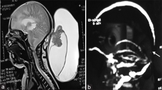
(a) Sagittal MRI of brain, there are two different sac with variegated intensity, (b) MR venography shows there is a gap between the sagittal and transverse sinus

(a) Sagittal MRI of brain, there are two different sac with variegated intensity, (b) MR venography shows there is a gap between the sagittal and transverse sinus