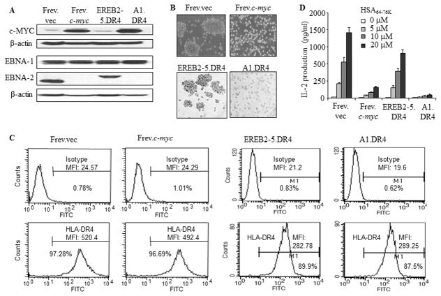
Figure 1.
Overexpression of c-MYC imparts a non-immunogenic BL phenotype to B-LCL and diminishes HLA-DR4-mediated CD4+ T cell recognition of BL-type cells. A, The wild-type B-cell line Frev which constitutively expresses HLA-DR4 molecules was either transfected with an empty vector or c-myc by electroporation. EREB2-5 cells are immortalized by EBV expressing a conditional estrogen receptor EBNA2 fusion protein (EREBNA2), and cellular proliferation is dependent on the availability of estrogen. A1 was established by stable transfection of conditionally EBV immortalized EREB2-5 cells with a c-myc/Igκ expression plasmid. The B-LCL-type cell line EREB2-5 and its derivative A1 (BL-type) were retrovirally transduced with HLA-DR4. Frev.vec, Frev.c-myc, EREB2-5.DR4 and A1.DR4 cells were then analyzed by western blotting for c-MYC, EBNA1, and EBNA2 proteins as described in the methods. β-actin was used as a loading control. B, Cells were subjected to microscopy for analysis of morphological characteristics (magnification × 20). C, Cells were stained for surface HLA-DR4 molecules using 359-F10 antibody and matched isotype control (IN-1), and analyzed by a flow cytometer as described. D, For T cell recognition assay, cells were incubated with different concentrations (0, 5, 10, 20μM) of a synthetic HSA64-76K peptide for 4h, washed and cocultured with the peptide-specific T cell hybridoma (17.9) for 24h. T cell production of IL-2 in the culture supernatant was quantitated by ELISA and expressed as pg/ml±SEM. Data are representative of three separate experiments.

