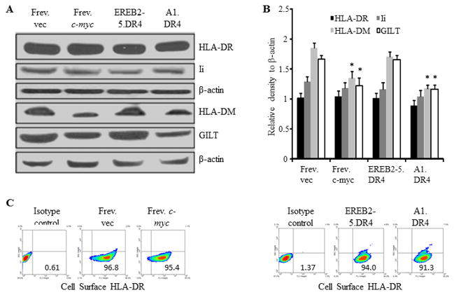
Figure 2.
Overexpression of c-MYC minimally influences HLA class II and Ii proteins, but alters other components of the class II pathway. A, B-LCL-type cells (Frev.vec and EREB2-5.DR4) and BL-type cells (Frev.c-myc and A1.DR4) were analyzed by western blotting for the expression of HLA-DR (L243), Ii (Pin 1.1), peptide editor HLA-DM and lysosomal thiol-reductase GILT (Santa Cruz antibodies) proteins. β-actin was used as a loading control. B, Densitometric analysis of protein bands detected in Figure 2A. Data are average density of three independent measurements of a representative band image and expressed as relative density (protein band/actin) ± S.D. Significant differences in relative band intensity were calculated by the Wilcoxon Rank Sum test; *p ≤0.0022. C, Flow cytometric analysis showing percent cell surface class II DR protein expression in BL (c-mychigh) and B-LCL (c-myclow) type cells. Frev.vec, Frev.c-myc, EREB2-5.DR4, and A1.DR4 cells were stained with an antibody against HLA-DR (L243), and a matched isotype (NN4) control, followed by flow cytometric analysis.

