
Figure 5.
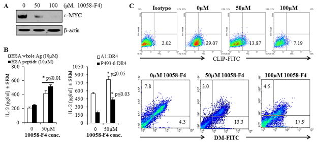
Treatment with the c-MYC inhibitor 10058-F4 partially restored CD4+ T cell recognition of c-MYC overexpressing P493-6 cells via the HLA class II pathway. P493-6 cells were grown in complete RPMI1640 medium in the absence of estrogen and tetracycline (BL-like phenotype). A, Expression of c-MYC proteins in P493-6 cells treated with various concentrations (0, 50 and 100μM) of the c-MYC inhibitor 10058-F4 for 24h. Cells were then washed and subjected to western blotting for analyzing c-MYC protein expression. β-actin was used as a loading control. B, left panel: P493-6 cells were cultured in the presence or absence of c-MYC inhibitor 10058-F4 (50μM) for 24h, followed by the addition of whole HSA or HSA64-76K peptide (10 μM) for the last 4h of incubation. Cells were washed, fixed with 1% paraformaldehyde, and cocultured with the HSA64-76K peptide specific T cell hybridoma (17.9) for 24h. T cell production of IL-2 in the culture supernatant was quantitated by ELISA and expressed as mean pg/ml±SEM. B, right panel: A1.DR4 and P493-6.DR4 cells which express endogenous Igκ were cultured in the presence or absence of c-MYC inhibitor 10058-F4 (50μM) for 24h. Cells were washed, fixed with 1% paraformaldehyde, and cocultured with the HLA-DR4-restricted Igκ188-203 peptide specific T cell hybridoma (2.18a) for 24h. Supernatants obtained were tested by ELISA to determine IL-2 levels as a measure of T-cell proliferation. Data are representative of three separate experiments. *p<0.05. C, Flow cytometric analysis of P493-6 cells treated with the c-MYC inhibitor 10058-F4 for 24h. Cells were first treated with 10058-F4 and stained with an antibody against CLIP (Cer-CLIP antibody) plus a matched isotype control (NN4 antibody) for determining surface CLIP protein expression (upper panel). Cells treated with 10058-F4 were also subjected to intracellular staining with antibodies against HLA-DM and HLA-DO molecules as described in the methods. The lower panel shows the effect of c-MYC inhibitor 10058-F4 on differential expression of HLA-DM and HLA-DO proteins in P493-6 cells as compared to untreated controls.