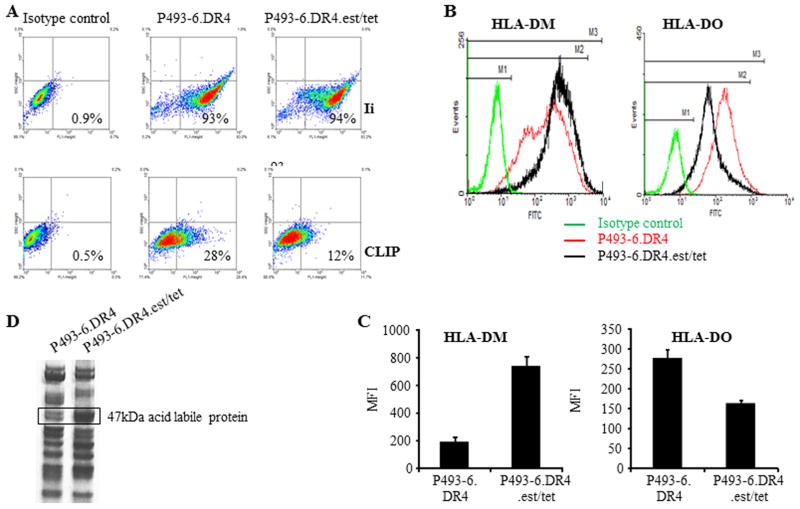
Figure 6.
Overexpression of c-MYC alters cell surface CLIP by regulating DM/DO ratio in BL/B-LCL type cells. A, DR4 expressing P493-6 cells were cultured under c-myc-on [(P493-6.DR4)(minus estrogen, minus tetracycline)] and c-myc-off conditions [(P493-6.DR4.est.tet)(plus estrogen, plus tetracycline). Cells were then intracellularly stained with antibody against Ii (Pin1.1), followed by addition of FITC-labeled secondary antibody as described. Cells were also stained with cer-CLIP antibody for cell surface CLIP proteins and analyzed by flow cytometry. B, P493-6 cells grown under c-myc-on and c-myc-off conditions were also intracellularly stained with antibodies against HLA-DM/HLA-DO proteins and appropriate isotype controls, followed by flow cytometric analysis. C, Bar graphs showing mean fluorescence intensity ±SEM of HLA-DM and HLA-DO staining in P493-6.DR4 and P493-6.DR4.est.tet cells. Data are representative of at least three separate experiments. *p<0.001. D, Analysis of a 47kDa protein differentially expressed in BL- vs B-LCL-type cells. Acid eluates obtained from BL-type (P493-6.DR4) and B-LCL-type (P493-6.DR4.est.tet) cells were separated and detected on a non-reducing gel as described in the methods. An approximately 47kDa band was excised and analyzed by MALDI-TOF/TOF mass spectrometry. Data shown are representative of at three separate experiments.

