
Figure 3.
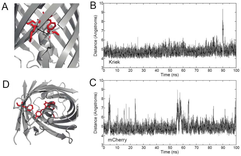
Comparative Molecular Dynamics of Kriek and mCherry. (A) Interstrand dynamics were quantitated by measuring the time-dependent distance between the α-carbons on A145 (β-7) and K198 (β-10), highlighted in red. (B) 100 ns time-trajectory of interstrand dynamics for Kriek and (C) mCherry. (D) Alternative 3O2 pathway, and ’gatekeeper’ residues, Q64, and F99, highlighted in red.