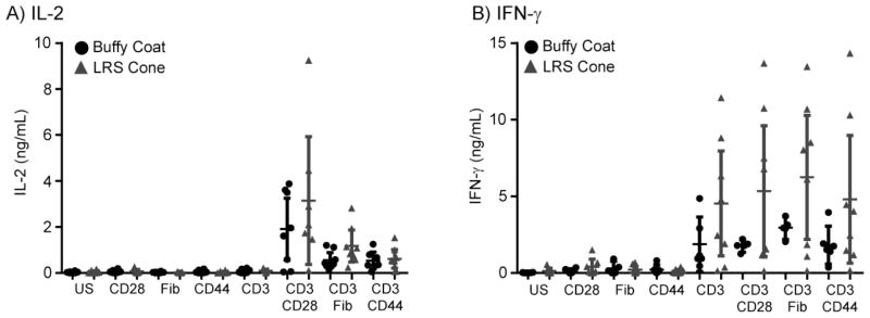
Figure 2.

Cytokine production and donor-to-donor variability is increased in the LRS cone isolated cells. T cells isolated from buffy coats or LRS cones were unstimulated (US) or stimulated with plate-bound anti-human CD3 (2 μg/ml) in the absence or presence of anti-CD28 (1 μg/ml), Fibronectin (1 μg/ml) or anti-CD44 (1 μg/ml) for 48 hours at 37 degrees. Supernatants were collected and assayed by ELISA for (A) IL-2 production and (B) IFN-γ production. The averages ± SEM of six different donors for each isolation method are shown with the outliers removed as described in the Materials and Methods.
