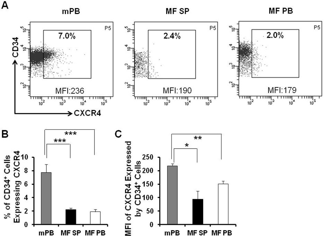
Figure 2. Down-regulated expression of CXCR4 by MF splenic and PB CD34+ cells.
(A) A representative flow cytometric plot demonstrating the proportion of mPB, splenic and PB MF CD34+ cells expressing CXCR4 and mean fluorescence intensity (MFI) of CXCR4 on these CD34+ cells. Data of Patient E are shown. (B-C) The proportion of both splenic and PB MF CD34+ cells expressing CXCR4 (B) and the MFI of CXCR4 expression on both splenic and PB MF CD34+ cells were significantly decreased as compared with mPB CD34+ cells. N=6. mPB: G-CSF mobilized peripheral blood; MF SP: myelofibrosis spleen; MF PB: myelofibrosis peripheral blood. *, P < 0.05; **, P < 0.01; ***, P < 0.001.

