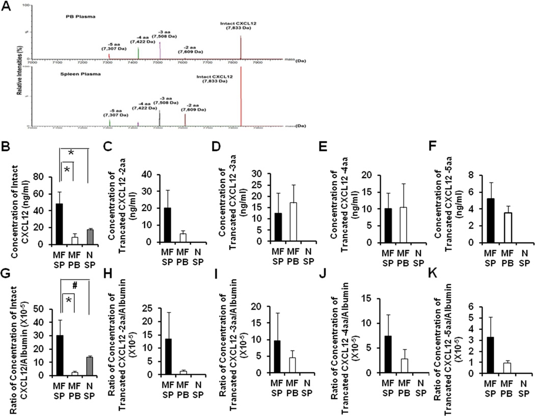
Figure 3. Concentrations of the intact CXCL12 and 4 truncated forms of CXC12 in splenic MF, PB MF and normal splenic plasma.
(A): The representative relative intensities of intact CXCL12 and four truncated forms in splenic (bottom) and PB plasma (top) of patient D. (B-F): Concentrations of intact CXCL12 (B) and four truncated forms (loss of 2 aa, 3 aa, 4 aa, and 5 aa) (C-F) of CXCL12 in paired splenic and PB MF plasma of all 6 patients and normal splenic plasma (n=7) were quantified using mass spectrometry. A higher level of intact CXCL12 is present in splenic MF plasma as compared with PB MF plasma or normal splenic plasma. The concentrations of the 4 truncated forms of CXCL12 were comparable in splenic and PB MF plasma, while none of the truncated forms of CXCL12 was detected in normal splenic plasma. (G-K): CXCL12 levels relative to albumin contained in corresponding splenic, PB MF and normal splenic plasma. The normalized level of intact CXCL12 in splenic MF plasma was again significantly higher than that of PB MF plasma (G). However, normal splenic plasma had marginally lower levels of intact CXCL12 than MF splenic plasma (G). The normalized levels of the 4 truncated forms of CXCL12 were found again similar in both splenic and PB MF plasma (H-K). *, P<0.05; #, P=0.08. aa: amino acid; MF SP: myelofibrosis splenic plasma; MF PB: myelofibrosis peripheral blood plasma; N SP: normal splenic plasma.

