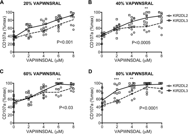
Figure 3.

Comparison of inhibition of CD158b+ NK cells from KIR2DL2+ and KIR2DL3+ donors to different combinations of VAP‐FA, VAP‐DA, and VAP‐RA. (A–D) 721.174 cells were incubated with combinations of three peptides as shown in Table 1 and used as target cells in degranulation assays for CD158b‐positive NK cells from KIR2DL2 (circles) and KIR2DL3 (squares) homozygous donors. The mean CD107a expression levels normalised to no peptide are connected by a dashed line (KIR2DL2 homozygous donors) or a full line (KIR2DL3 homozygous donors). The ratios of VAP‐RA to VAP‐FA were: (A) 20% VAP‐RA:80% VAP‐FA, (B) 40% VAP‐RA:60% VAP‐FA, (C) 60% VAP‐RA:40% VAP‐FA, and (D) 80% VAP‐RA:20% VAP‐FA. In all experiments, the VAP‐RA:VAP‐FA ratio was kept constant but the total quantity was varied in combination with VAP‐DA in order to maintain a total peptide concentration of 10 μM in the peptide mix (Supporting Information Table 1). Data show the mean value for each individual donor, and are from one experiment performed in duplicate. p values for the differences between the donors as determined by ANOVA are shown. For comparisons at individual peptide concentrations *p < 0.05, **p < 0.01, ***p < 0.001.
