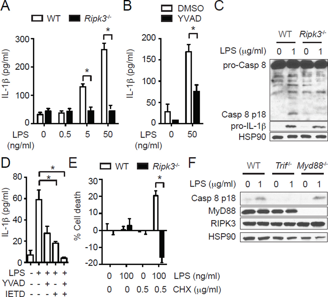
FIGURE 1.
RIPK3 promotes caspase 1- and caspase 8-dependent IL-1β secretion in BMDCs. (A) RIPK3 is required for LPS-induced IL-1β secretion in BMDCs. IL-1β secretion by WT and Ripk3−/− BMDCs stimulated with LPS for 6 hours was determined by ELISA (n=4). (B) Caspase 1 is partially responsible for RIPK3-dependent IL-1β secretion. WT BMDCs were pretreated with 10 µM z-YVAD-fmk (YVAD) for 1 hour and stimulated with LPS for 4 hours (n=4). IL-1β secretion was determined by ELISA. (C) RIPK3 is critical for LPS-induced caspase 8 activation. Cell lysates from WT and Ripk3−/− BMDCs stimulated with LPS for 1 hour were subjected to western blot analysis. (D) Caspase 1 and caspase 8 cooperate to mediate optimal IL-1β secretion. WT BMDCs pretreated with 10 µM YVAD and/or 10 µM z-IETD-fmk (IETD) for 1 hour were stimulated with 50 ng/ml LPS for 6 hours (n=4). IL-1β secretion was determined by ELISA. (E) Inhibition of protein synthesis converts the LPS-induced RIPK3 signal to one that causes apoptosis. WT and Ripk3−/− BMDCs pretreated with CHX for 1 hour were stimulated with LPS for 14 hours. Cell death was determined by measuring intracellular ATP level (n=3). (F) RIPK3-dependent caspase 8 activation requires an intact TRIF. Cell lysates from BMDCs of the indicated genotypes stimulated with LPS for 1 hour were subjected to western blot analysis. Results shown are mean ± SEM. Asterisks: p < 0.05.

