Figure 1.
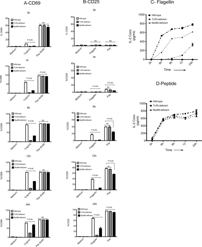
TLR5 is essential for activation of flagellin-specific T cells at low dose of flagellin. Time kinetics was performed to evaluate the activation of flagellin-specific SM1 T cells (CD4+CD90.1+) in vitro, using CD69 and CD25 as markers of activation. CD11c+ DCs were purified from spleens of WT (white bars), TLR5-deficient (gray bars), and MyD88-deficient DCs (black bars), using DC enrichment kits and were cultured in 1:1 ratio with flagellin-specific SM1 T cells in the presence of flagellin (1 ng/mL), peptide (6 μM), and medium alone for 3, 6, 9, 12, and 24 h. (A and B) The activation of flagellin-specific T cells as determined by (A) CD69 expression and (B) CD25 expression at given time points, was measured by flow cytometry. Data are shown as mean + SEM of three samples per group, and are from one experiment, representative of two independent experiments. (C and D) Concentration of IL-2 in DC culture supernatants at 3, 6, 9, 12, and 24 h, as measured by ELISA. CD11c+ DCs purified from spleens of WT (filled circles), TLR5-deficient (filled squares), and MyD88-deficient DCs (filled triangles) using DC enrichment kits and were cultured in 1:1 ratio with flagellin-specific SM1 T cells in the presence of (C) flagellin (1 ng/mL) or (D) peptide (6 μM) for 3, 6, 9, 12, and 24 h. Data are shown as mean ± SEM of three samples per group carried out in tissue culture replicates, and are from one single experiment representative of two independent experiments. NS: nonsignificant by unpaired t-test.

