Figure 4.

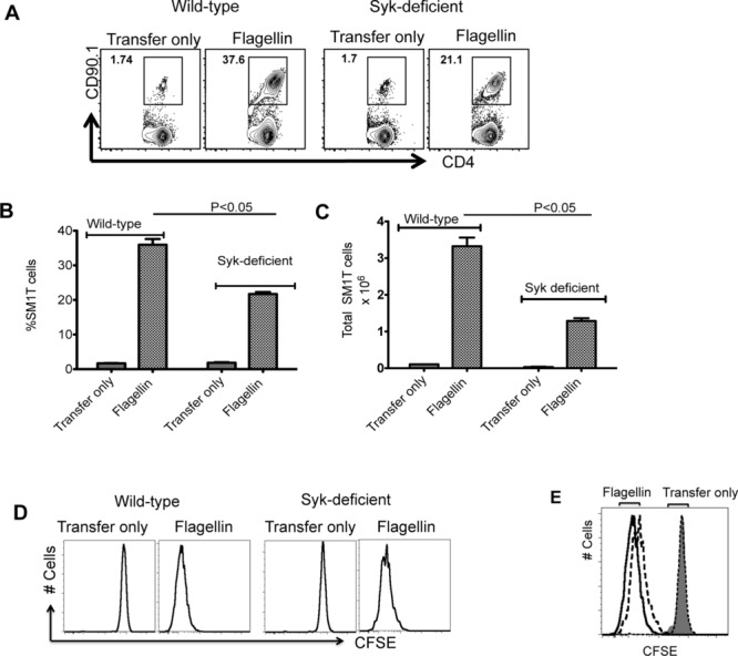
Syk deficiency hinders robust activation of SM1 T cells in vivo. (A) In total, 1 × 10⁶ CFSE-stained SM1 T cells were adoptively transferred into WT and Syk-deficient chimeric mice and the following day were immunized with 1 μg of flagellin. Three days later, clonal expansion of SM1 T cells was examined in the spleen by flow cytometry. Representative flow cytometry plots (n = 3 mice per group) are shown. (B and C) WT or Syk-deficient mice were immunized with flagellin (1 μg) and (B) the percentage and (C) total number of SM1 T cells in the spleens was determined by flow cytometry. Data are shown as mean + SEM of three mice per group, and are from a single experiment representative of three independent experiments. NS: nonsignificant by unpaired t-test. (D and E) SM1 T-cell proliferation in WT and Syk-deficient mice was examined by CFSE dye dilution. (E) WT (solid line), Syk-deficient mice (dotted line). Shaded area shows isotype control staining. Representative flow cytometry plots (n = 3 mice per group) are shown and are from one single experiment representative of three independent experiments.
