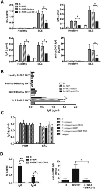
Fig. 4. Expanded iNKT cells from SLE patients, but not healthy subjects, can induce IgG production.

(A) B cells were cultured for 10 days with expanded autologous iNKT cells from healthy subjects (n=5) or lupus patients (n=5) in the presence or absence of anti-CD1d or isotype control antibody. Immunoglobulin in the culture supernatants was measured by ELISA. (B) B cells from healthy subjects were co-cultured with autologous iNKT cells or iNKT cells from SLE patients for 10 days in the presence of anti-CD1d or isotype control antibody. Alternatively, B cells from SLE patients were co-cultured with autologous iNKT cells or iNKT cells from healthy donors. IgG in the supernatant was measured by ELISA. (C) B cells from SLE patients were stimulated with PWM and SAC in the presence or absence of anti-CD1d, anti-iNKT (6B11) or anti-iNKT TCR (TCRVα24/TCRVβ11) antibodies for 10 days. (D) To assess the ability of iNKT cells from lupus patients to induce Ig class switching, sorted IgM+CD27-CD19+ naïve B cells were cultured with autologous iNKT cells for 10 days in the presence or absence of anti-CD1d antibody. Antibodies in the supernatant were measured by ELISA. Left panel shows total IgG and IgM, and right panel shows anti-dsDNA IgG. Antibodies in the supernatant were measured by ELISA. (A) Data are shown as mean ± SD of five samples pooled from five independent experiments. (B-D) Data are shown as mean ± SD of three samples pooled from three independent experiments. * indicates P<0.05, ** indicates P<0.01. (Paired t-test used).
