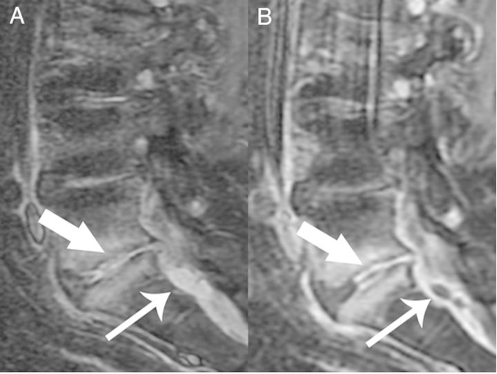
Figure 1.
Sagittal T1 Short T1 Inversion Recovery (A) and postcontrast T1 fat-saturated (B) images demonstrate confluent epidural enhancement with mass effect upon the thecal sac. There are also two small T2 hyperintense, rim-enhancing fluid collections consistent with small epidural abscesses (thin arrows). In addition, there is increased T2 signal and enhancement in the L5-S1disc space consistent with discitis-osteomyelitis (thick arrow).

