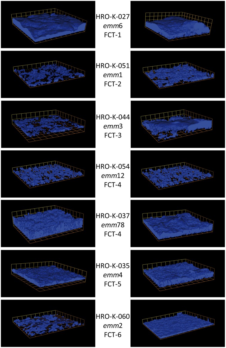
Figure 1.
Three-dimensional images of biofilms of various GAS emm/FCT types grown in C-medium in the absence or presence of glucose. Cells were stained with Alexa Fluor 647 and visualized via Confocal Laser Scanning Microscopy (CLSM); magnification 600 times; box size 13.8 × 13.8 μm. Left panel: non-supplemented C-medium. Right panel: C-Medium supplemented with 30 mM glucose. Strain names, emm-types, and FCT types are given in the middle column.

