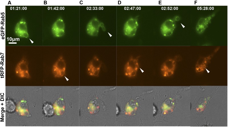
FIG 3 .
Sequential localization of Rab5 and Rab7 during live-cell imaging of phagosome maturation. Shown are RAW264.7 macrophages coexpressing eGFP-Rab5 and tRFP-Rab7. Transfected macrophages were incubated with the C. albicans Δefg1 mutant (CA79), and the temporal kinetics of Rab5 and Rab7 localization to individual phagosomes were observed at 1-min intervals. Selected frames are shown with times indicated in the upper panels (h:min:s). White arrowheads indicate the localization of Rab5 to newly engulfed C. albicans cells (upper row), with sequential loss of the Rab5 signal and acquisition of the Rab7 signal (middle panels), which occurs throughout the movie, during which several fungal particles are sequentially engulfed. The corresponding movie is available in the supplemental material (see Movie S5). The scale is shown by a white bar.

