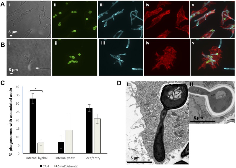
FIG 6 .
Diminished actin polymerization around phagosomes containing hyphae of the C. albicans Δmnt1/2 mutant imaged in 3D. RAW264.7 cells were combined with live C. albicans cells from strain CAI4 (NGY152) (A) or the Δmnt1/2 mutant (NGY337) (B) in standard phagocytosis assays for 2 h prior to fixing of cells, staining, and imaging. The scale is shown by white bars. Representative DIC (i) and composite z-stack (ii to v) images are shown: extended focus (images ii to v) is shown following phagocytosis of both strains. Cells were stained as follows: prephagocytosis FITC staining of C. albicans cells (ii), postfix CFW staining of fungal cell wall chitin (iii), postfix macrophage phalloidin staining of actin (iv), and merge (v). (C) Quantification of polymerized actin associated with phagosomes containing C. albicans determined from analysis of >90 phagosomes (% association with internalized hyphal or yeast parts of C. albicans phagosomes or with entry or exit points of the macrophage). *, P ≤ 0.05. (D) Differential phagosomal membrane apposition surrounding yeast and hyphal parts of C. albicans wild-type strain CAI4 within J774.1 (i) and RAW264.7 (ii) cells visualized by TEM.

