FIG 4 .
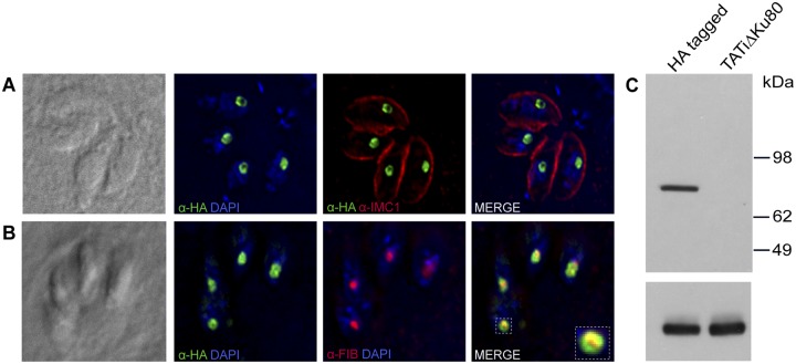
Localization and expression of the T. gondii SUN protein. (A) Fluorescence microscopy of the C-terminally HA-tagged SUN protein using anti-HA (green) and IMC1 (red [to outline parasite cells]) antibodies shows its nucleolar localization. Nuclei are stained with DAPI (4′,6-diamidino-2-phenylindole) (blue). (B) SUN protein is associated with the granular compartment (outer) of the nucleolus. Fibrillarin antibody staining of the dense fibrillar compartment is shown in red, and the HA-tagged SUN protein (green) forms a ring-shaped structure around the fibrillarin. (C) Western blot of parasite pellets showing expression of HA3-tagged SUN protein using anti-HA antibody. The TATi ΔKu80 parental line showing no expression is included as a control. The lower panel shows the loading control using anti-α-tubulin antibody.

