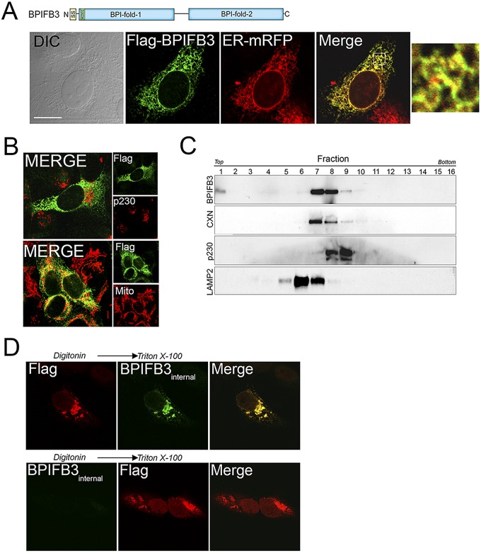
FIG 2 .
Localization of BPIFB3 to the ER. (A, top) Schematic of BPIFB3. (Bottom) Confocal microscopy for Flag (green) and ER-mRFP (red) in U2OS cells transfected with BPIFB3-Flag and infected with CellLights ER-RFP baculovirus. (Left) Differential interference contrast (DIC) image. (Right) A 5× magnification of the area indicated by the white box in the merged image. (B) Confocal microscopy for Flag (green) and either p230/Golgi (red, top) or MTCO2 (to label mitochondria; red, bottom) in U2OS cells transfected with BPIFB3-Flag at ~48 h posttransfection. (C) Subcellular fractionation of BPIFB3 in U2OS cells stably expressing BPIFB3-Flag. Shown are immunoblots from collected fractions for BPIFB3-Flag, calnexin (CXN), p230/Golgin, and LAMP2. (D) Confocal microscopy of U2OS cells transiently transfected with BPIFB3-Flag. At 48 h posttransfection, cells were permeabilized with digitonin, fixed with PFA, and then incubated with anti-Flag-M2 (top row) or BPIFB3 (bottom row) antibodies. Cells were then permeabilized with Triton X-100 and stained with anti-BPIFB3 (top row) or anti-Flag M2 (bottom row) antibodies. Bar, 10 µm.

