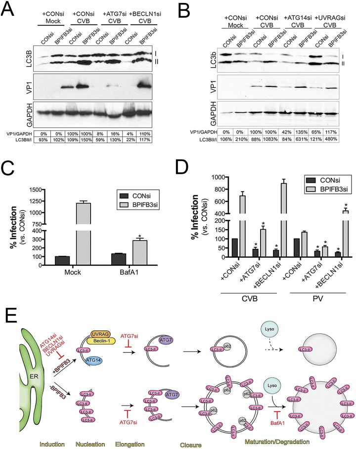
FIG 7 .
Silencing of BPIFB3 enhances CVB infection by the induction of autophagy, independent of the core autophagy initiation machinery. (A) Immunoblots for LC3B (top), VP1 (middle), and GAPDH (bottom) from HBMEC transfected with CONsi or BPIFB3si, cotransfected with either control, ATG7, or beclin-1 (BECLN1) siRNAs, and then infected with CVB (1 PFU/cell). Nonlipidated (I) and lipdated AP-associated LC3B (II) results are shown. At bottom, densitometry analysis for VP1 (normalized to GAPDH results and presented as the percent change from CONsi-transfected levels for either CONsi or BPIFB3si) and the percentage of LC3B-II normalized to LC3B-I. (B) Immunoblots for LC3B (top), VP1 (middle), and GAPDH (bottom) from HBMEC transfected with CONsi or BPIFB3si, cotransfected with either control, ATG14, of UVRAG siRNAs, and then infected with CVB (1 PFU/cell). Nonlipidated (I) and lipdated AP-associated LC3B (II) results are shown. At the bottom, densitometry analysis results for VP1 (normalized to GAPDH and presented as the percent change from CONsi-transfected levels for either CONsi or BPIFB3si) and the percentage of LC3B-II normalized to LC3B-I are shown. (C) HBMEC transfected with CONsi or BPIFB3si were infected with CVB in the absence (mock) or presence of BafA1, and then infection was assessed by RT-qPCR. Data were normalized to mock-treated conditions in CONsi-transfected cells. (D) HBMEC transfected with CONsi or BPIFB3si and either CONsi, ATG7si, or beclin-1 siRNAs were infected with PV (1 PFU/cell) or CVB (1 PFU/cell) for ~18 h, and infection was assessed by RT-qPCR. Data in panels C and D are means ± standard deviations. *, P < 0.01. (E) Schematic for the mechanism by which BPIFB3 regulates CVB replication. At top, in cells expressing BPIFB3, CVB replication induces autophagy and requires the expression/activity of core components of the autophagy initiation machinery (ATG14, UVRAG, beclin-1) and a component involved in elongation (ATG7). Hatched lines indicate that it is unknown if CVB elicits autophagic flux. Below, in cells with low levels of BPIFB3, CVB infection proceeds in the absence of the core initiation machinery, enhancing the levels of LC3B-II, promoting autophagic flux, and enhancing the sizes of replication organelles.

