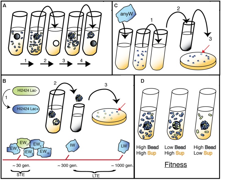
FIGURE 1.
Bead model of biofilm evolution. (A) Bead-transfer method used in both evolution experiments. (1) A fresh bead is added to an inoculated tube containing marked (long-term evolution, LTE) or both marked and unmarked (short-term evolution, STE) WT populations and grown for 24 h. (2) Bead is colonized by biofilm cells and transferred to a fresh tube containing a sterile bead and growth media. (3) Bacteria disperse from original bead and attach to new bead. (4) New bead is selected and process repeats; note that 48-h bead is archived to sample representative population. (B) (1) Lac+ strain used in LTE and STE experiments, Lac- used in STE only. (2) Following daily transfer of colonized bead, remaining 48 h bead was removed and vortexed in PBS. (3) Populations were plated and screened for different morphologies related to adaptive traits (red arrow indicates a novel W mutant). Early Wrinkly (EW) mutants were detected at ∼30 generations, while Intermediate Wrinkly (IW) and Late Wrinkly (LW) were detected at ∼300 and 1000 generations. (C) Isolation of Smooth mutants. 1. W colonies were serially passaged in planktonic environment for three days. (2) Samples were diluted and plated. (3) Smooth colonies were detected and isolated. (D) Predicted fitness effects of adaptive mutants (blue cells) in sup and bead environment, with three outcomes presented: from left to right, early colonization and rapid dispersal; rapid dispersal but delayed colonization; early colonization and slow dispersal.

