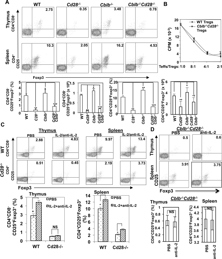
FIGURE 1.
Loss of Cbl-b partially rescues defective development of CD4+CD25+Foxp3+ Tregs in Cd28−/− mice. (A) Thymocytes and splenocytes from WT, Cblb−/−, Cd28−/−, and Cblb−/−Cd28−/− mice (n=5/group; 8 wks of age) were surface-stained with anti-CD4, anti-CD8, and anti-CD25, and intracellularly stained with anti-Foxp3. The percentage of CD4+CD8−CD25+Foxp3+ cells in thymuses and CD4+CD25+Foxp3+ cells in spleens of different groups of mice were calculated. Statistical significance was calculated using the student t-test. *p<0.05 compared to WT, ** p<0.01 compared to WT; #p<0.001 compared to Cd28−/−. (B) WT CD4+CD25− T cells were incubated with CD4+CD25+ T cells isolated from WT and Cblb−/−Cd28−/− mice at different ratios in the presence of anti-CD3 and T-depleted APCs for 72 hrs. T cell proliferation was determined by [3H]thymidine incorporation. (C) WT and Cd28−/− mice received daily i.v. injections of a combination of mouse IL-2 (1.5 µg/day, eBioscience) and anti-mouse IL-2 mAb (clone JES6-1A12) (50 µg/day; BioXCell) for 7–9 days. Thymuses and spleens were analyzed for Tregs on day 8–10 by flow cytometry as above. (D) Cblb−/−Cd28−/− mice were treated with anti-IL-2 daily by i.v. for 7–9 days, and Tregs in thymuses and spleens were analyzed by flow cytometry. *p<0.05 compared to PBS-treated controls; NS, no significance. The data shown are one representative of three independent experiments.

