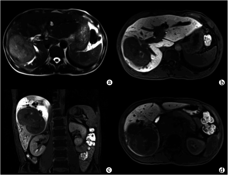
Figure 1.

Exophytic peripheral cholangiocarcinoma in a patient with abdominal pain. A T2-weighted image (a) showed a large, hyperintense mass with lobulated margins in the right hepatic lobe, and the adjacent bile duct was dilated. The hepatocellular phase (b, c) showed that the tumor was inhomogeneously hypointense compared with the liver parenchyma. The liver parenchyma also showed inhomogeneous enhancement on the hepatocellular phase (c). The biliary phase (d) showed that the common bile duct was completely filled with Gd-EOB-DTPA, indicating that the function of biliary system was normal.
