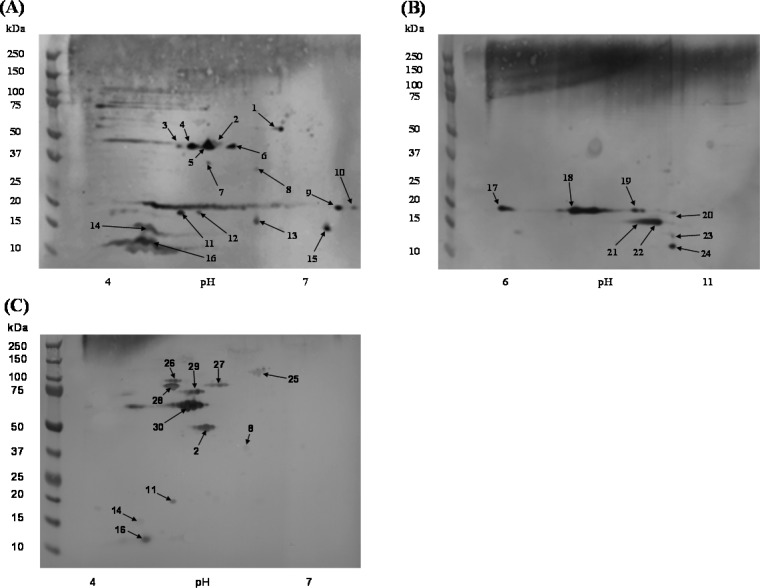
Figure 2.
2D Western blot of MPF and F. tularensis LVS immune sera to MPF proteins. (A) MPF resolved in a pH range of 4–7 and probed with MPF immune sera. (B) MPF resolved in a pH range of 6–11 and probed with MPF immune sera. (C) MPF resolved in a pH range of 4–7 and probed with F. tularensis LVS immune sera. The numbered arrows correspond to the spots labeled in Figure 1 and the protein identifications presented in Table 1 and Table S2 in the Supporting Information.

