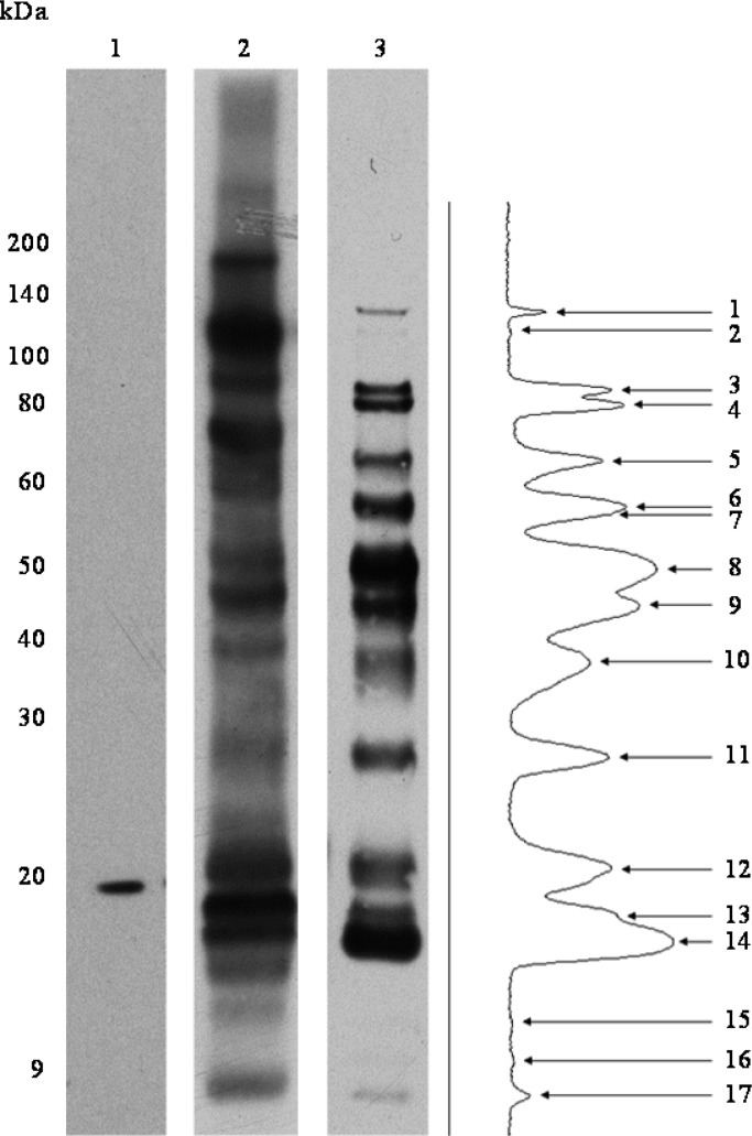
Figure 3.
Biotinylation of F. tularensis LVS surface-exposed proteins. Antibiotin Western blots to detect biotinylated proteins after labeling F. tularensis LVS surface proteins with LC-Biotin. Lane 1, WCL of F. tularensis LVS unlabeled control; Lane 2, labeled F. tularensis LVS WCL (15 s exposure); Lane 3, labeled F. tularensis LVS intact cells (2 min exposure) accompanied by densitometry analysis of reactive protein bands.

