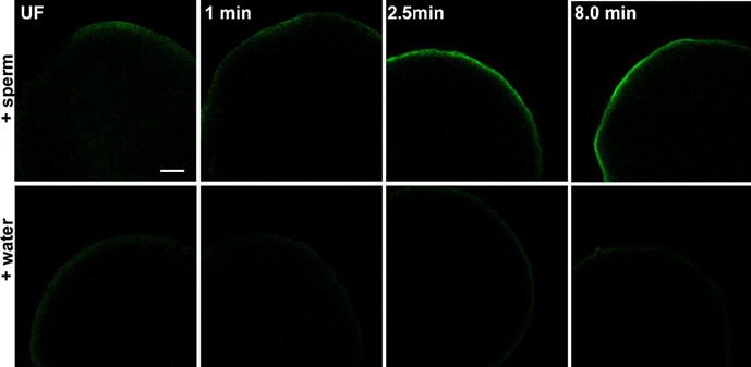
Figure 8. Response to activation by sperm or hypotonic conditions.

Eggs were fixed for immunofluorescence analysis before (UF) or at 60 seconds, 2.5 minutes, or eight minutes after activation by sperm and water (+sperm) or water alone (+water). Immunolabeling with the clone 28 antibody was performed after chorion removal and conditions of illumination and signal amplification in the fluorescence channel were maintained at a constant level to allow comparison of immunofluorescence intensity. All zygotes were oriented with the animal pole toward the top of each panel. Magnification is indicated by the bar, which represents 100μM.
