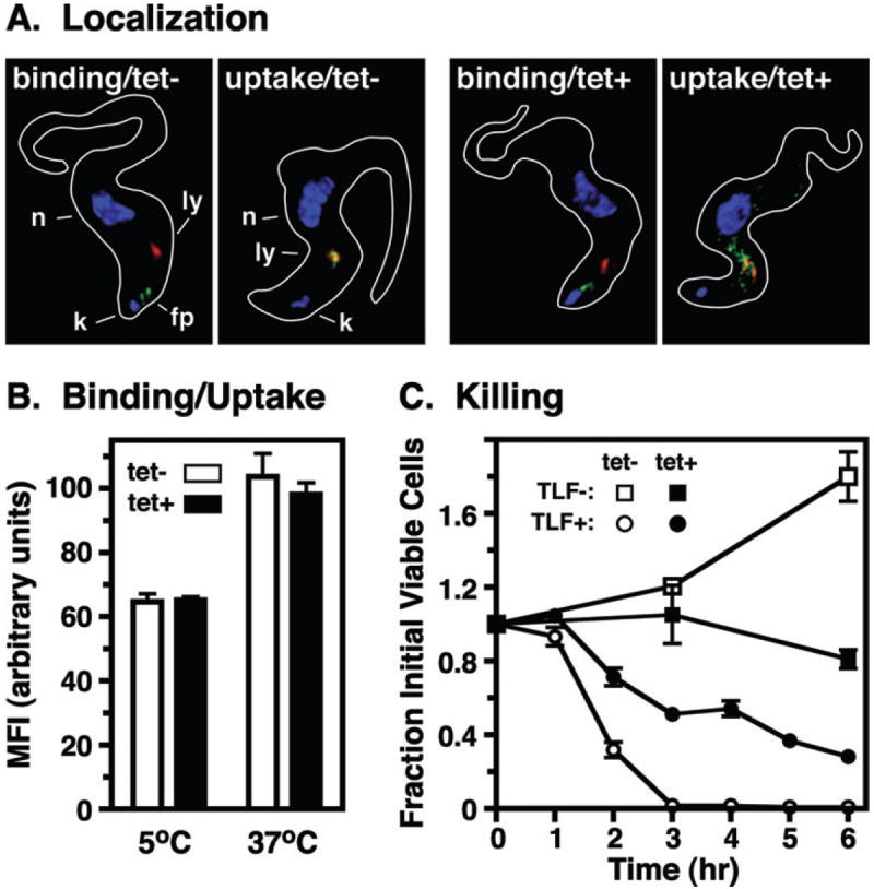
Fig. 7.

Effect of TbRab7 silencing on TLF killing. Control (tet−) and TbRab7 silenced (tet+) BSF cells were incubated with purified TLF. Cells were incubated with TLF:A488 for flagellar pocket binding and uptake as in Fig. 4.
A. For microscopy cells were fixed/permeabilized and stained with anti-p67 (red) and TLF:A488 was detected directly (green). The positions of nuclei (n), kinetoplasts (k), lysosomes (ly), and flagellar pockets (fp) are indicated (control only). B. Cell associated fluorescence in fixed cells was quantitatively assessed by flow cytometry. Mean fluorescent intensities (MFI) are presented (means ± SEM, n = 6). Unpaired t-tests indicated no significant differences (P > 0.5) between control vs. silenced cells for both binding and uptake. C. TLF killing. Cells were incubated at 37°C with or without unconjugated TLF. Cell death was scored by phase microscopy and is presented over time as fraction of initial viable cells (means ± SEM, n = 3). Rate of killing was determined as the slope (m) following regression analysis over the linear phase (control, 1–3 h, m = 45.7%/h, r2 = 0.9440; silenced, 1–6 h, m = 13.8%/h, r2 = 0.8562).
