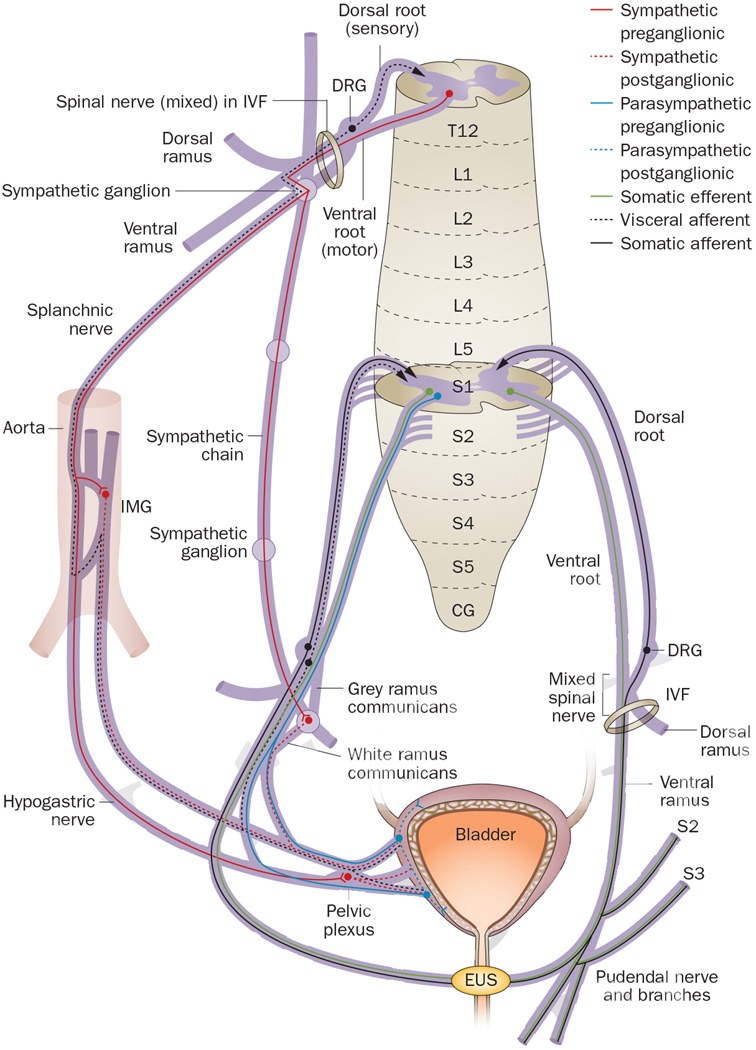
Figure 1.
Spinal cord neuroanatomy and bladder innervation in humans and other mammals. After exiting the spinal cord, dorsal and ventral roots (which consist of small rootlets that unite to form the large root) join into a mixed spinal nerve that divides into a dorsal ramus, a ventral ramus, the sympathetic chain, and splanchnic nerves. The bladder is innervated via sympathetic axons that project from T10 to L2 (only T12 contributions are illustrated) through either splanchnic nerves to the inferior mesenteric ganglion on the aorta or descend within the sympathetic chain to upper sacral ganglia, where they synapse on neurons that project to the bladder. Parasympathetic bladder innervation from pelvic splanchnic nerves consists of preganglionic axon projections from S2 to S4 (only S2 contributions are illustrated) in most mammalian species to ganglia located near the bladder wall. Sensory feedback travels to the spinal cord from visceral and somatic structures by ‘hitchhiking’ on autonomic and somatic motor nerves. Abbreviations: CG, coccygeal segment; DRG, dorsal root ganglion; EUS, external urethral sphincter; IMG, inferior or caudal mesenteric ganglion; IVF, intervertebral foramen; L, lumbar; S, sacral; T, thoracic.

