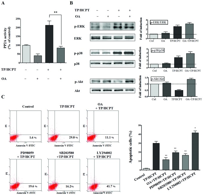
Figure 6.
Effect of PP2A inhibition on ERK, p38 and Akt signaling pathways, as well as TP/HCPT-induced apoptosis. (A) TP/HCPT-enhanced PP2A activity was inhibited by OA. A549 cells were also treated with OA (50 nM) for 2 h prior to 24 h of combinatorial treatment with TP (25 ng/ml) and HCPT (4 μg/ml). Cell lysates were prepared and assayed for phosphatase activity of PP2A. The values are expressed as a percentage of the control (untreated cells; 100% PP2A activity). **p<0.01 vs. the TP/HCPT group (n=3). (B) Effect of PP2A inhibition on ERK, p38 and Akt signaling pathways. Cell lysates after different treatments were prepared and subjected to western blot analysis. The results are representative of three independent experiments, and the corresponding densitometric analyses for relative protein expression were shown in the right hand panels. (C) Effect of inhibitors of pp2A, p38, ERK and Akt on TP/HCPT-induced apoptosis. The cells were treated with OA (50 nM), p38 inhibitor-SB 203580 (10 μM), ERK inhibitor-PD 98059 (10 μM) and Akt inhibitor-LY294002 (25 μM) for 2 h prior to 24 h of TP/HCPT treatment. Then the cells were collected and analyzed using Annexin V/PI double staining, and bar graphs shown in the right hand panel representing the apoptotic rates. *p<0.05 and **p<0.01 vs. the TP/HCPT group (n=3).

