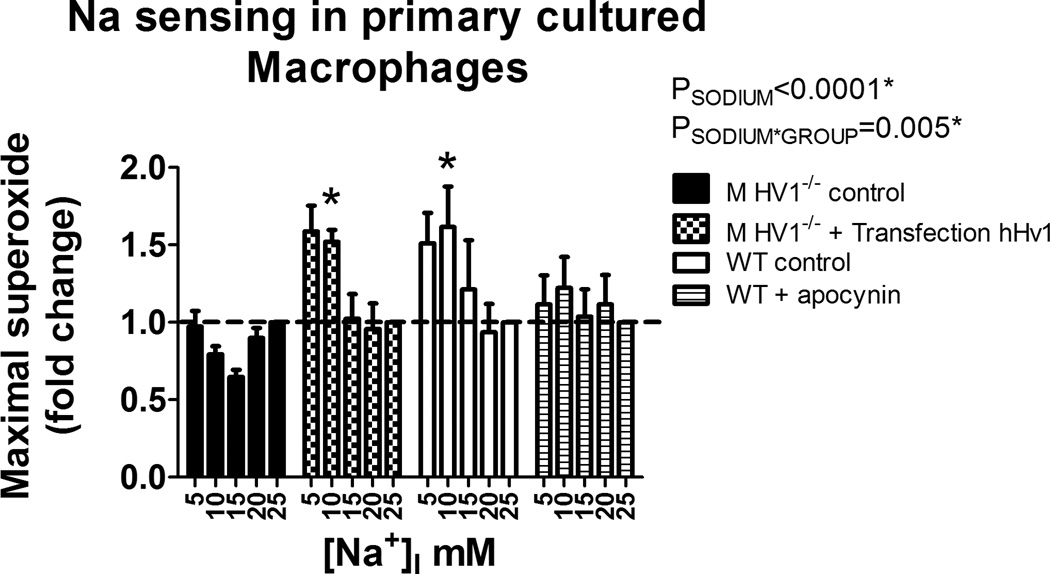
Figure 5. Contribution of Hv1 and NAD(P)H oxidase toward Na sensing in primary cultured macrophages.
X-axis, intracellular Na+ concentration ([Na+]I). Y-axis, maximal superoxide production in freshly isolated peritoneal rat macrophages following addition of PMA (100mM) determined by L-012 luminescence (data normalized to the response at 25mM [Na+]I within each animal). HV1−/− control (closed columns; n=5); Wild-type (WT) control (open columns; n=5); Wild-type + apocynin 100µM (striped columns; n=5); HV1−/− transfected with human Hv1 (checked columns; n=5). Data were compared using 2-way ANOVA with Bonferroni post-hoc test comparing HV1−/− control to all other groups. * = p<0.05. Data are mean±SE.

