
Figure 1.
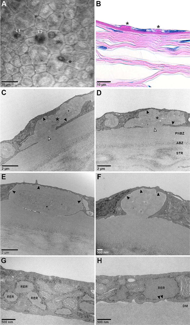
Varying appearances of guttae in homozygous Q455 K mutant mouse corneas. (A) Corneal confocal microscopy shows endothelial cell polymegathism and pleomorphism. Multiple forms of guttae are present in close proximity including Laing et al’s stages 1 (L1) and 2 (L2), and a distinct, sharply raised gutta suggested by Gottsch et al to be a characteristic of late onset FECD (*). (B) Periodic acid Schiff staining shows differently sized guttae (*). (C,D) Transmission electron microscopy shows large, irregularly shaped masses of material (*) with similar electron density and quality as the posterior non-banded zone (PNBZ) of Descemet membrane (DM) which appear to be intracellular as there is no obvious separation with the adjacent cytoplasm (closed arrowheads). Also shown is the anterior banded zone (ABZ) of DM adjacent to the stroma (STR). Internal features suggestive of cellular structures are present as are areas of focal attachment to DM (open arrowheads). (E,F) The other major form of this material shows more regular borders, more homogeneous internal features, and areas of clear separation from the cytoplasm (closed arrowheads). (G) Endothelial cells show areas of markedly dilated rough endoplasmic reticulum (RER). (H) In some areas the dilated RER becomes closely approximated to the basal cell membrane with loss of ribosomes (arrowheads) suggesting potential areas of membrane fusion which would enable contact and attachment between DM and RER contents. These areas of attachment could become the stalks of guttae.