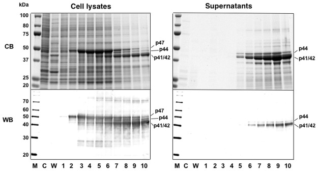
Fig 1. Time course of expression of MCPyV-VP1.

Insect Tn5 cells were infected with AcMCPyV-VP1 and harvested for the indicated time periods. Five microliters of culture medium and samples of lysate from 105 cells were analyzed by SDS-PAGE. Protein bands were visualized by Coomassie blue staining (CB; upper panels) and Western blotting with anti-MCPyV VP1 antiserum (WB; lower panel). M, molecular weight marker; C, mock-infected; W, wild-type baculovirus-infected cells; lanes 1 to 10, 1 to 10 days p.i.
