
Figure 3.
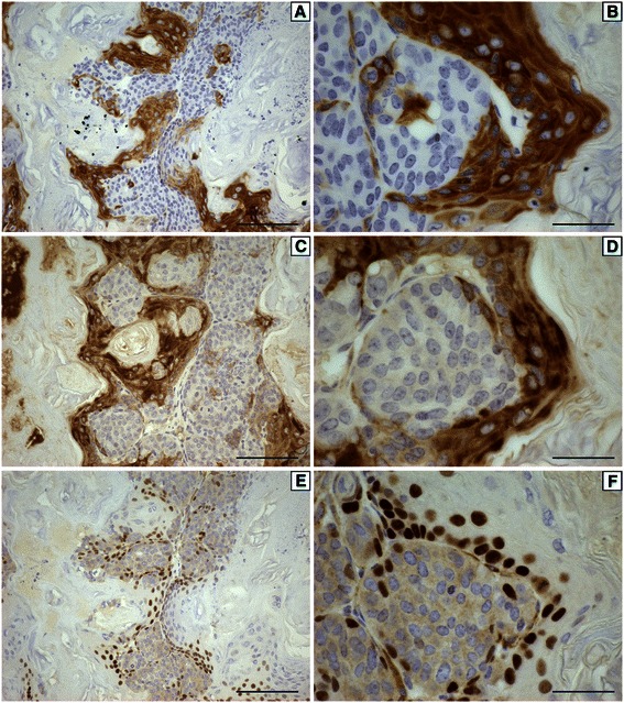
Representative immunohistochemistry for cytokeratin 5 (A,B), cytokeratin 14 (C,D) and p63 (E,F) from a mammary tumor that developed in a soy-fed mouse following IGF-IR induction at 100 days of age at 200x (A,C,E) or 600x (B,D,F) magnification. Scale bars in A,C,E are 100 μM while scale bars in B,D,F are 33 μM.