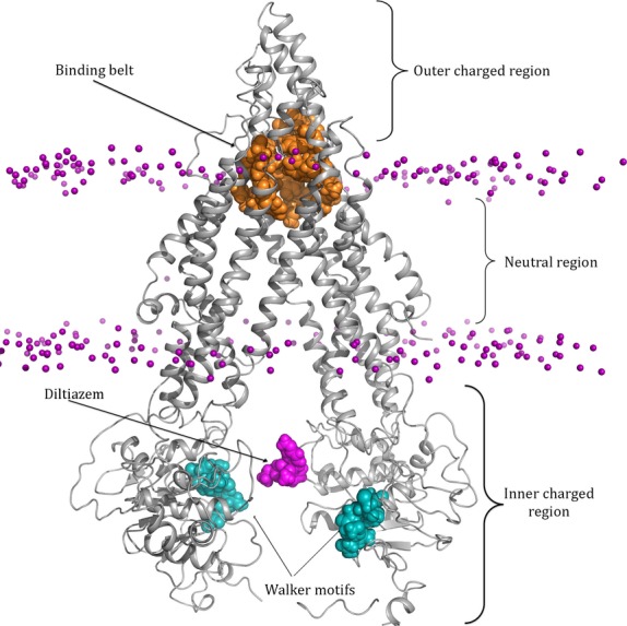
Figure 1.

Structure of P-glycoprotein placed in a POPC phospholipid membrane. Pgp is shown in cartoon representation and colored gray. The phosphate heads are shown in sphere representation and colored purple. Pgp has three distinct regions: the charged extracellular matrix (ECM) region, the neutral lipid domain, and the charged cytoplasmic region. Conserved ATP-binding domains Walker A and Walker B are shown in spheres and colored teal. The drug-binding belt is represented by the residues colored orange that start at the boarder of the neutral lipid domain and extend into the charged ECM region. The drug Diltiazem colored magenta has been placed between the NBDs of Pgp demonstrating the initial drug placement for the cytoplasmic recruitment study. The lipid tails and water molecules are not shown. POPC, 1-palmitoyl-2-oleoyl-sn-glycerol-3-phosphocholine.
