
Figure 1. Detection of FFA2 using RK1101 antibody in rat tissues.
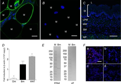
Whole mount mesenteric white adipose tissue or a cryostat section of oesophagus was incubated with anti-FFA2 antibody RK1101 (green) with or without blocking peptide, counterstained with 4′,6-diamidino-2-phenylindole (DAPI, blue). Mesenteric adipocytes were positively stained (A), whereas pre-absorption abolished the staining (B). ld, lipid droplet. Internal bars: 20 μm. C, oesophageal mucosa, consisting of stratum layers (St), lamina propria mucosae (LPM), muscularis mucosae (MM) and submucosa (SM), was negatively stained, with faint staining observed in the muscularis propria (MP). L, lumen. Internal bars: 100 μm. D, expression of FFA2 in oesophageal mucosa (Om), duodenal bulb mucosa (Bm) and white adipose tissue (WAT) assessed by real time PCR with the ΔCt method. Each data point represents the mean ± SEM (n = 6 rats). *P < 0.05 vs. Bm. E, Western blot for FFA2 using RK1101 in duodenal mucosa (Bm) (left panel; FFA2) and pre-absorption with blocking peptide (right panel; + P). M, molecular marker with size (kDa) on the left. F, FFA2-transfected cells (a) were positively stained with RK1101 (red), whereas FFA2-transfected cells were negatively stained with pre-absorbed antibody (b). FFA3-transfected cells (c) and mock-transfected cells (d) were negatively stained with RK1101. Counterstained with DAPI (blue). Internal bars: 100 μm.