
Figure 2. Localisation of FFARs in rat duodenum.
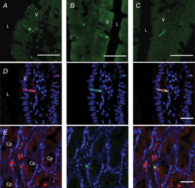
Frozen cryostat sections were incubated with the primary antibody for FFA1 (A), FFA2 (B) or FFA3 (C). L, lumen; V, villus. Internal bars: 100 μm. D, double staining with FFA2 (red; RK1101 antibody, left), 5-HT (green; middle) and merged image (right). Counterstained with DAPI (blue). L, lumen; V, villus. Internal bars: 20 μm. E, double staining with FFA3 (red; left), GLP-1 (green; middle) and merged image (right). Counterstained with DAPI (blue). Cp, crypt. Internal bars: 20 μm.