


Figure 7. AKT activation is involved in ILK protection against uraemic toxin-induced apoptosis of EA.hy926 cells.
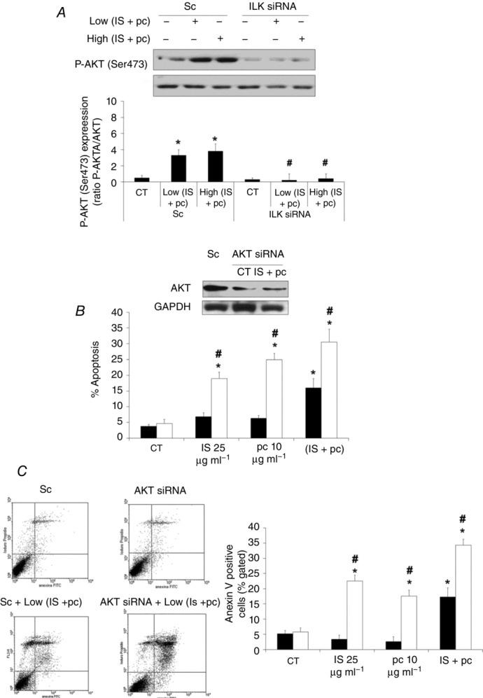
A, cells were depleted of ILK with specific siRNA (100 nm) and afterwards incubated in medium supplemented with 2.5% normal serum (NS) plus a combination of low concentrations of indoxyl sulfate (25 μg ml−1) and p-cresol (10 μg ml−1) (Low IS + pc) or plus a combination of high concentrations of IS (100 μg ml−1) and pc (100 μg ml−1) (High IS + pc) for 24 h. Scrambled RNA (Sc) was used as control. Representative Western blots of phosphorylated AKT in the serine-473 residue (P-AKT (Ser473)) and total AKT as control are shown. Bars represent the normalized densitometric analysis of blots against control values. B and C, cells were depleted of AKT with specific siRNA (open bars) (100 nm) and treated afterwards with IS (25 μg ml−1), pc (10 μg ml−1) or both, for 24 h. Scrambled RNA (Sc) (filled bars) was used as control. After incubation, apoptosis was determined in PI-stained cells and analysed by flow cytometry (B) or cells were stained with annexin V-FITC and PI, followed by flow cytometry analysis (C). Some results are shown by representative dot plots. Data are expressed as mean ± SEM of five independent experiments. *P < 0.05 vs. control (CT; 2.5% NS, 24 h); #P < 0.05 vs. Sc.