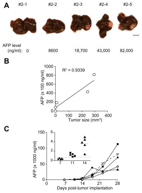
Fig. 2. Orthotopically implanted human HCC tumor model.
HCC tumors were surgically implanted into the liver of SCID mice. (A) Macroscopic view of the implanted tumor developed in the liver of a representative SCID mouse with their corresponding serum AFP level indicated below the picture. Scale bar = 5 mm. (B) AFP levels plotted against tumor size. R2 = 0.9339 and p = 0.01. (C) Growth kinetics of the orthotopically implanted HCC tumor. Tumor growth increased rapidly when serum AFP level was >500 ng/ml, indicated by the dotted line (in the inset). (This figure appears in colour on the web.)

