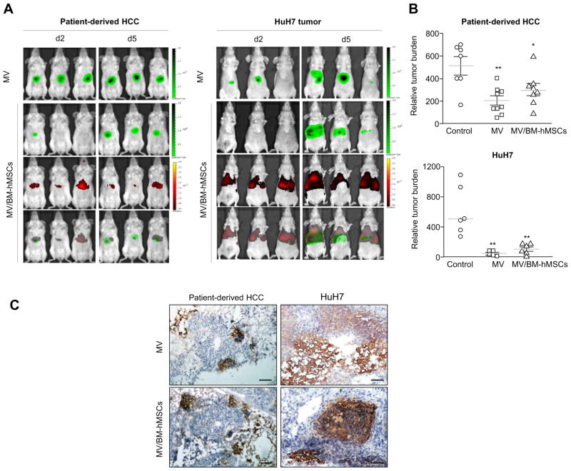
Fig. 3. Systemic delivery of MV infection via infected BM-hMSCs to orthotopically implanted HCC tumors resulted in significant tumor growth suppression.
(A) Tumor-bearing mice were given 2 × 10⁶ TCID50 MV-Luc or 2 × 10⁶ MV-Luc-infected DiR-labeled BM-hMSCs (MV/BM-hMSCs). Bioluminescence imaging of virally encoded firefly luciferase is shown in green, and fluorescence imaging of DiR-labeled BM-hMSCs is indicated in red. Overlaid images (bottom panel) showed colocalization of both signals indicating delivery of MV-Luc infection (green) by infected BM-hMSCs (red). (B) Relative tumor burden at necropsy; **p <0.01; *p <0.05. (C) Immunohistochemistry staining for MV-N protein. Scale bar = 50 μm. (This figure appears in colour on the web.)

