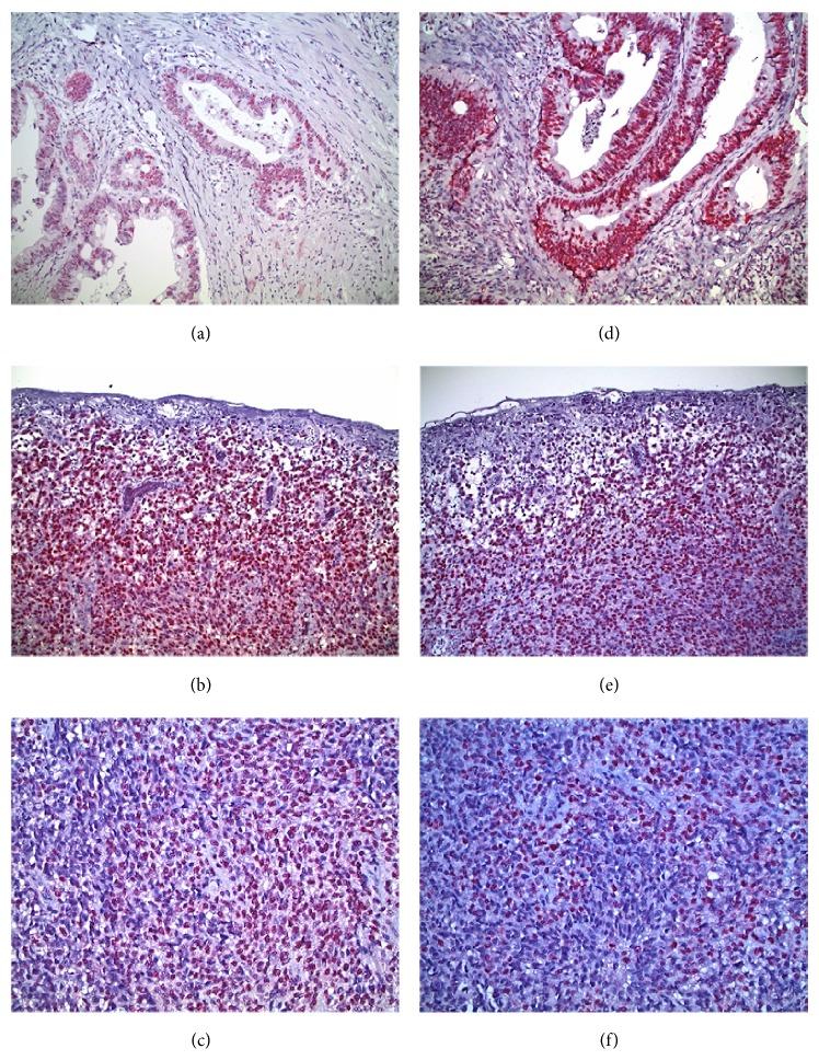
Figure 2.
Immunohistochemistry analysis for the expression of the hMLH1 and hMSH2 MMR proteins in mucosal melanomas. (a) MMR-proficient/BRAF mutant colorectal cancer control exhibiting positive nuclear staining for hMLH1. (b) Vertical growth phase mucosal melanoma with ulcerated surface. Almost all melanoma cells display intense nuclear staining for hMLH1. (c) Mucosal melanoma with spindle and epithelioid cells strongly positive for hMLH1. (d) MMR-proficient/BRAF mutant colorectal cancer control displaying positive nuclear staining for hMSH2. (e) Vertical growth phase mucosal melanoma with ulcerated surface and diffuse nuclear staining for hMSH2. (f) Mucosal melanoma with spindle and epithelioid cells strongly positive for hMSH2. Photomicrographs (a), (b), (d), and (e) are of ×20 magnification, and photomicrographs (c) and (f) are of ×40 magnification.

